Fixing the Roof While Furnishing the House: Do CXL and Speciality Contact Lenses Complement or Compete in the Management of Keratoconus?

With the increasing use of corneal cross-linking (CXL) for keratoconus, some have questioned whether this treatment could reduce the demand for specialised contact lenses.  

Keratoconus is a well-known condition among practitioners that fit speciality contact lenses. This progressive disorder affects approximately 1 in 500 individuals and is much more common than previously assumed .1,2 The severity of this corneal ectasia varies, often presenting asymmetrically between the eyes.3 It is characterised by corneal thinning, reduced best-corrected visual acuity and significant refractive changes over a short period of time. Eventually, irregularity in corneal shape leads to visual distortions that cannot always be corrected with conventional optical aids such as glasses or soft contact lenses.3 

This is where speciality contact lenses, particularly rigid gas-permeable (RGP) lenses of different designs, play a crucial role. These lenses create a smooth refractive surface, compensating for corneal irregularities. This reduces higher-order aberrations and often restores functional vision where other methods, such as glasses and soft lenses, fail.4 As a result, speciality contact lenses are often the only viable solution for many keratoconus patients, enabling them to function effectively in both their professional and personal lives. But why would we be content with smacking a RGP lens on an unstable cornea? Contact lenses do not treat the underlying condition, meaning that corneal irregularity can continue to worsen, potentially reaching the point where a transplant is required.

Let’s start by fixing the roof!

Since the FDA’s approval of the CXL treatment for keratoconus in 2016,5 the number of corneal transplants has drastically decreased.2 A decade ago, corneal transplantation was the standard procedure for these patients. Today, it is considered a last resort.6 To simplify the concept of CXL, one can think of it as a process that accelerates the stiffening that occurs naturally in the corneal stroma, the thickest layer of the cornea, as we get older. It is this stiffening which explains why keratoconic progression typically slows with age.3 The goal of CXL is to strengthen the corneal stroma by applying riboflavin (vitamin B2) and UVA light, thereby preventing further ectatic progression.7 The combination of riboflavin and UV-A induces the formation of free oxygen radicals, facilitating the development of additional covalent bonds within the stromal collagen. This strengthening process improves the cornea’s biomechanical and biochemical stability, playing a vital role in managing keratoconus.8 CXL is highly effective, with studies demonstrating a reduction in ectasia progression of up to 70%.9 The procedure is generally recognised as safe, with a minimal risk of complications. However, while most side effects are temporary and reversible, certain complications may occur, including corneal haze and scarring, a decline in both uncorrected and best spectacle-corrected visual acuity, infectious and non-infectious keratitis, stromal melting, and, in some cases, treatment failure with continued ectasia progression.7 These factors must also be considered when determining the appropriate treatment for mild keratoconus, particularly in children and young adults. In clinical practice, the approach tends to be that if there is progression of ectasia and the eye can tolerate CXL, treatment is considered. This is regardless of the current level of visual impairment. The reasoning behind this is that stabilizing the cornea first provides a foundation for future optical correction. By doing so, the natural cornea can be preserved and the need for a corneal transplant may be reduced. In essence, CXL is aimed at halting disease progression rather than improving vision directly. 
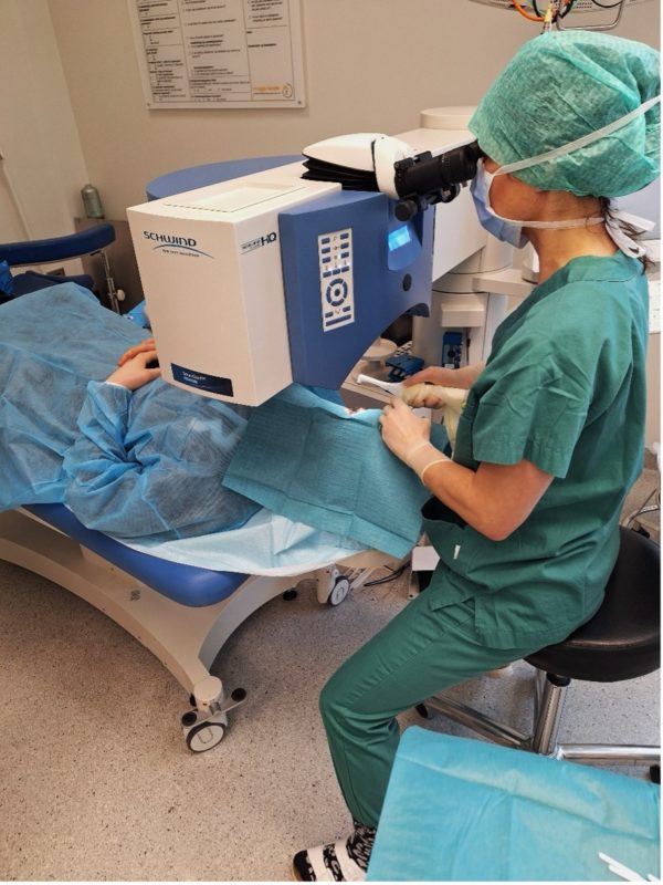
Removal of a patient’s epithelium with an Amoils brush prior to CXL treatment 

So now that the roof is fixed. What about the furniture?

When choosing vision correction for keratoconus, both corneal RGP and scleral lenses offer unique benefits. Corneal RGP lenses, a long-standing option for keratoconic eyes, provide sharp vision but may be challenging for some patients to tolerate. These lenses often provide excellent visual acuity and are particularly effective in correcting regular astigmatism. In cases of more severe pathology, where RGP lenses may become unstable on-eye, scleral lenses offer an alternative. These larger RGP lenses rest on the sclera, creating a fluid-filled reservoir that protects the cornea while ensuring stability, comfort, and improved vision, especially for irregular or ectatic corneas.4 The best choice depends on individual needs and disease severity. Studies confirm that scleral lenses contribute to functional improvement in patients with keratoconus, particularly in more advanced cases. Additionally, findings suggest that these lenses play a significant role in enhancing quality of life, regardless of disease severity or the extent of visual improvement.10,11 
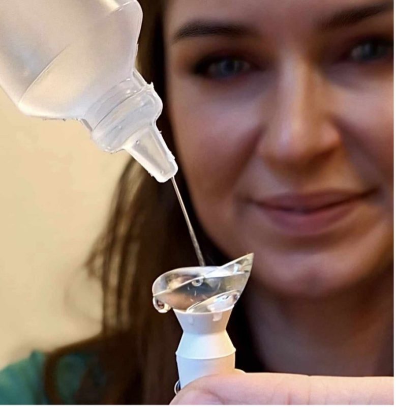
Modern scleral lenses are highly tuneable to wearers needs, making them ideal for keratoconic eyes 

Could CXL fully replace contact lenses for keratoconic patients?

Let us, just for a moment, play with the idea that we would truly try to use CXL to undermine the speciality contact lens industry. To achieve this, we would firstly need to screen every 10-15-year-old across the globe using advanced and costly Scheimpflug technology. Upon diagnosis, we would have to CXL treat each and every one of them, ensuring that progression of keratoconus is halted before it could ever lead to the need for specialised lenses. Ethical considerations relating to age and the risk of complications would also have to be considered. Doesn’t sound very realistic, does it? 

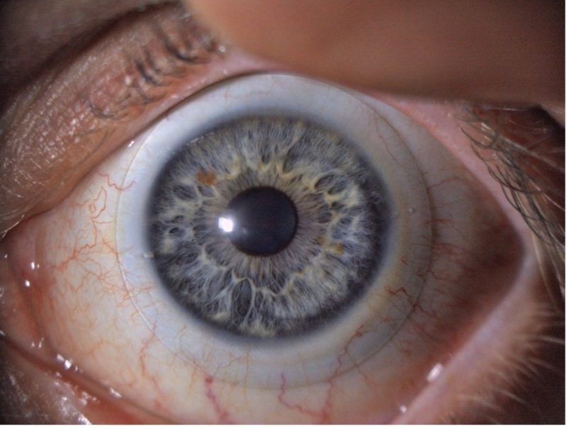
A keratoconic eye with scleral lens in place 

Conclusion: We must address the fundamental clinical challenges while simultaneously optimising visual outcomes

Effective management of keratoconus is not just about halting disease progression but also about ensuring that patients achieve the best possible vision. CXL plays a crucial role in stabilising the cornea, but it does not eliminate the need for precise optical correction. In fact, one could argue that without the rapid advancements in specialised contact lenses, CXL would not have been the success it is today. By combining structural stabilisation with advanced visual rehabilitation, we move beyond simply managing the disease, we actively enhance the patient’s quality of life. Because what is the point of fixing the roof only to live in a house without furniture?


References

1. Godefrooij DA, de Wit GA, Uiterwaal CS, Imhof SM, Wisse RP. Age-specific Incidence and Prevalence of Keratoconus: A Nationwide Registration Study. Am J Ophthalmol. 2017;175:169-72.
2. Kristianslund O, Hagem AM, Thorsrud A, Drolsum Prevalence and incidence of keratoconus in Norway: a nationwide register study. Acta Ophthalmol. 2021;99(5):e694-e9.
3. Santodomingo-Rubido J, Carracedo G, Suzaki A, Villa-Collar C, Vincent SJ, Wolffsohn JS. Keratoconus: An updated review. Cont Lens Anterior Eye. 2022;45(3):101559.
4. van der Worp E. A Guide to Scleral Lens Fitting (2 ed.). 2. ed: Scleral Lens Education Society; 2015. p. 19.
5. Gomes JA, Tan D, Rapuano CJ, Belin MW, Ambrósio R, Jr., Guell JL, et al. Global consensus on keratoconus and ectatic diseases. Cornea. 2015;34(4):359-69.
6. Matthaei M, Sandhaeger H, Hermel M, Adler W, Jun AS, Cursiefen C, et al. Changing Indications in Penetrating Keratoplasty: A Systematic Review of 34 Years of Global Reporting. Transplantation. 2017;101(6):1387-99.
7. Agarwal R, Jain P, Arora R. Complications of corneal collagen cross-linking. Indian J Ophthalmol. 2022;70(5):1466-74.
8. Naranjo A, Manche EE. A comprehensive review on corneal crosslinking. Taiwan J Ophthalmol. 2024;14(1):44-9.
9. Wollensak G, Spoerl E, Seiler T. Riboflavin/ultraviolet-a-induced collagen crosslinking for the treatment of keratoconus. Am J Ophthalmol. 2003;135(5):620-7.
10. Baudin F, Chemaly A, Arnould L, Barrénéchea E, Lestable L, Bron AM, et al. Quality-of-Life Improvement After Scleral Lens Fitting in Patients With Keratoconus. Eye Contact Lens. 2021;47(9):520-5.
11. Kreps EO, Pesudovs K, Claerhout I, Koppen C. Mini-Scleral Lenses Improve Vision-Related Quality of Life in Keratoconus. Cornea. 2021;40(7):859-64.

Author spotlight

Pia Victoria Haugum Ekker

Pia graduated with a Master’s degree from the University of South-Eastern Norway and worked in private practice for over a decade. Her current role, as a specialist optometrist at St. Olavs Hospital in Trondheim, combines all aspects of contact lens fitting with performing corneal cross-linking surgeries.