Optimising Ocular Health in Ectodermal Dysplasia: A Case Series on Tailored Scleral Lens Fitting

Background:

Ectodermal Dysplasias (ED) are genetic conditions affecting the development of skin, hair, teeth, nails, and sweat glands1. Ocular findings include lacrimal drainage obstruction and hypoplasia, punctal agenesis, distichiasis, trichiasis, meibomian gland alterations and ankyloblepharon with sequelae of chronic dry eye2,3.

Complications include chronic ocular pain, neurotrophic keratitis, and corneal opacification4. Among other treatments, scleral lenses (SL) can optimize vision and maintain the health of the ocular surface. This case series highlights two patients with ED successfully fit with large diameter SLs.

Case 1:

42-year-old Caucasian female presented for dry eye and scleral lens consultation. H/o ED with malformation of nasolacrimal ducts OU, herpes zoster keratitis OU in 2017 with corneal scarring OS > OD. Habitual scleral lens use with inadequate fit due to corneal touch.

Quadrant specific SLs were designed with a larger diameter and increased central clearance, to be used in conjunction with autologous serum tears to improve the ocular surface. After 2 months of wear, corneal punctate staining fully resolved.

Figures 1a and 1b: Slit lamp examination of the cornea OD/OS: Mixed NaFl staining secondary to lens touch. Corneal irregularity due to central corneal scarring, thinning, and neovascularization. Diffuse bulbar conjunctival staining secondary to exposure keratopathy.
Figures 2a and 2b: View of anterior segment OD/OS: Sparse lashes of upper and lower eyelids, inflamed lid margins and bulbar conjunctiva, and subepithelial scarring of the central cornea with accompanied thinning.
Figures 3a and 3b: Slit lamp view of scleral lenses OD/OS with optic section demonstrating adequate fluid reservoir clearance over areas of scarring and thinning.
4a:  640 µm of clearance over area of scarring and irregularity.
4b:  204 µm of clearance over area of scarring and irregularity.
Figures 4a and 4b: AS-OCT of final lens design.
 

Type

Power

BC

Diameter

Total Sag

Edges by Quadrant

Material

OD

BostonSight

FSE1

-16.61

7.98

17.0

2455

1: +150

2: STD

3: +150

4: +150

Optimum Extreme

OS

BostonSight

FSE1

-7.39

9.72

17.0

2300

1: STD

2: STD

3: STD

4: STD

Optimum Extra

Table 1: Final scleral lens design OD and OS

Case 2:

56-year-old Caucasian male presented with eye irritation and blurred vision. H/o of EEC syndrome (ectrodactyly, ED, and orofacial clefts)

Ocular history of limbal stem cell deficiency, keratoconjunctivitis sicca and severe primary open angle glaucoma. Patient reported eye pain OD>OS while using habitual SLs. Impressions were obtained. Custom, impression-based SLs improved patient comfort and reduced ocular inflammation after 1 month of wear.

Figures 5a and 5b: Initial appearance OD/OS: Irregular/thickened lid margins with telangiectasia, diffuse conjunctival injection, and peripheral corneal stromal haze with neovascularization involving the visual axis.
 
Figures 6a and 6b: View of OD/OS after 1-year of SL wear: Reduced ocular inflammation and regression of corneal neovascularization without surgical intervention.
 

Type

Power

BC

Diameter

Back Optic Zone Diameter

Material

OD

EyeFitPRO

-3.75

8.058

16.5

9.24 x 7.85

Optimum Extreme with HydraPEG

OS

EyeFitPRO

-7.75

7.541

16.0

10.17 x 8.07

Optimum Extreme with HydraPEG

Table 2: Final scleral lens design OD and OS

Conclusions

This case series demonstrates the benefit of customized SL fitting with increased diameter to protect the cornea and conjunctiva of patients with ED. Improved SL fit and concomitant use of autologous serum can prevent further corneal scarring.

Both patients benefited from advanced SL customization which improved comfort and wear time. Scleral lenses effectively address multiple concurrent ocular issues, and achieving an accurate fit can significantly enhance long-term comfort, wear duration, and overall quality of life.

References

1. Landau Prat D, Katowitz WR, Strong A, et al. Ocular manifestations of ectodermal dysplasia. Orphanet Journal of Rare Diseases 2021; 16: 197.
2. Jen M, Nallasamy S. Ocular manifestations of genetic skin disorders. Clinical Dermatology 2016; 34(2): 242-75.
3. Keklikci U, Yavuz I, Tunik S, et al. Ophthalmic manifestations in patients with ectodermal dysplasia syndromes. Advances in Clinical and Experimental Medicine 2014; 23(4): 605-10.
4. Zhu L, Zhu H. Ocular herpes: the pathophysiology, management and treatment of herpetic eye diseases. Virologica Sinica. 2014; 29(6): 327-42.
5. Hassan OM, Farooq AV, Soin K, et al. Management of Corneal Scarring Secondary to Herpes Zoster Keratitis. Cornea 2017; 36(8): 1018-1023.

Author spotlight

Diana Masolak

Dr. Diana Masolak is a cornea and contact lens resident at the Illinois College of Optometry (ICO), with an emphasis in ocular disease. She graduated from the Illinois College of Optometry in 2024. She is from Downers Grove, Illinois, and attended the University of Illinois at Urbana-Champaign for her undergraduate studies. She is passionate about improving the functionality of her patients’ vision while increasing their ocular comfort, as well as preparing her students for life as independent clinicians. Her clinical interests include the fitting of specialty contact lenses for corneal ectasia, and myopia control.

Ellen Shorter

Dr. Ellen Shorter earned her Doctor of Optometry from Illinois College of Optometry in Chicago, graduating summa cum laude. After graduation, she has competed a residency in ocular disease and low vision at the Jesse Brown VA Medical Center and Hines Veteran’s Hospital, a PROSE clinical fellowship at the Boston Foundation for Sight and a Scholars for Teaching Excellence Faculty Fellowship at the University of Illinois.  


She is a fellow of both the American Academy of Optometry and the Scleral Lens Education Society.  Currently, Dr. Shorter is an Associate Professor of Ophthalmology at the Illinois Eye and Ear Infirmary specializing in medically necessary contact lenses.  

Jennifer Harthan

Dr. Jennifer Harthan is a graduate of the Illinois College of Optometry (ICO). After completing a Residency in Cornea and Contact Lenses at ICO, she became a full-time faculty member. Dr. Harthan is a Professor at ICO and Chief of the Cornea Center for Clinical Excellence at the Illinois Eye Institute. Dr. Harthan is a Fellow of the American Academy of Optometry, Scleral Lens Education Society and serves on the Medical Advisory Board for the International Keratoconus Academy. Dr. Harthan is a founding member of the SCOPE (Scleral Lenses in Current Ophthalmic Practice Evaluation) research team. She has numerous publications on the topics of complex contact lens fits and anterior segment disease. Dr. Harthan is actively involved in ocular surface disease and contact lens research and lectures on these topics at national and international meetings.

Katie Kwan

Dr. Kwan was born and raised in Canada, where she completed her undergraduate studies before earning her Doctor of Optometry degree from Illinois College of Optometry in Chicago, Illinois. After graduation, completed a residency in pediatric optometry at the Southern College of Optometry in Memphis, Tennessee. Currently, Dr. Kwan is pursuing a fellowship in medically necessary specialty contact lenses at the University of Illinois Chicago.

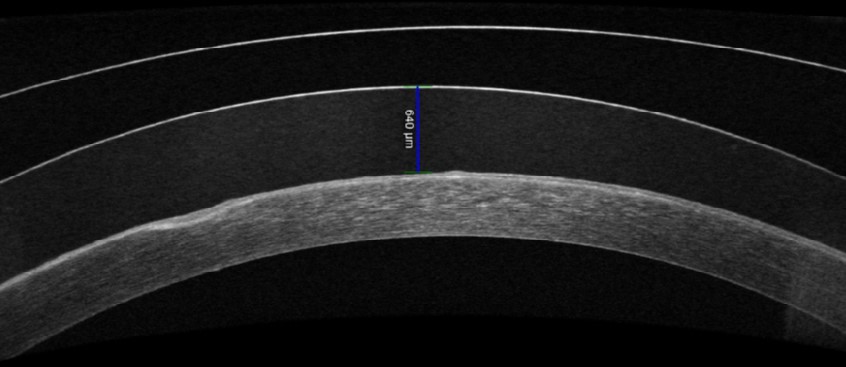
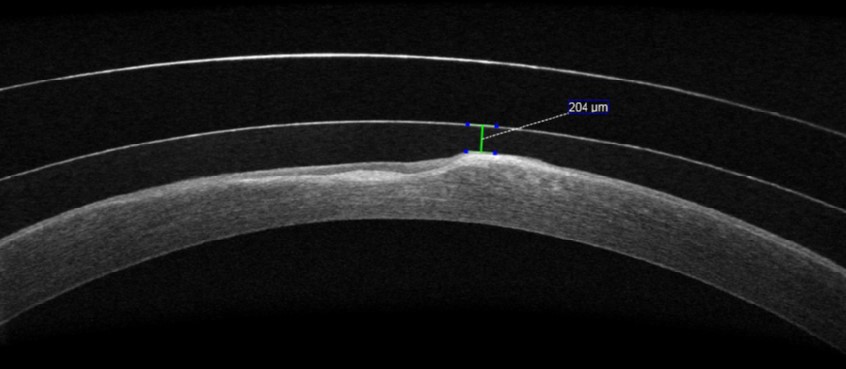
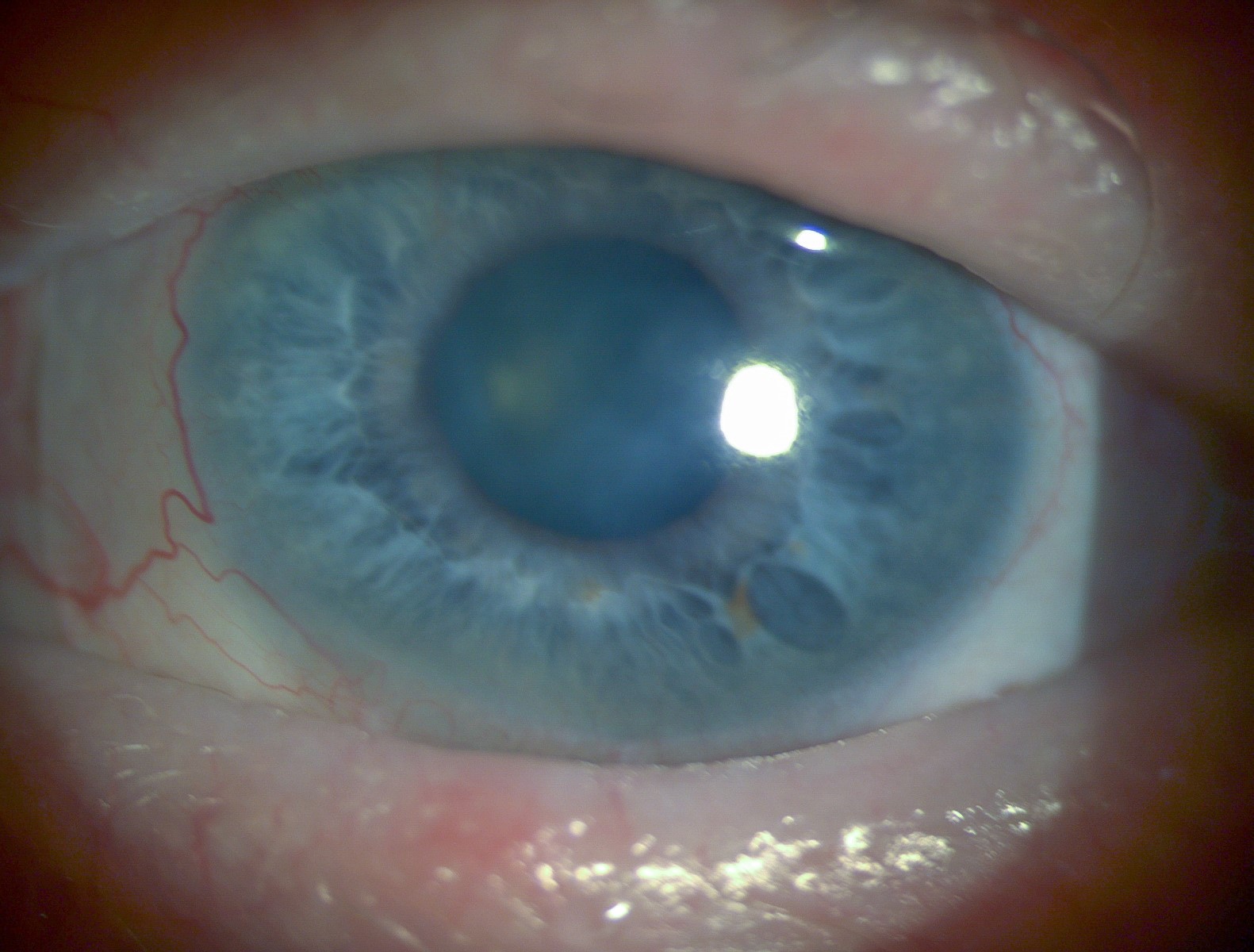
Her clinical interests include pediatric eye care, the management of ocular surface disease with medically necessary contacts and myopia control.