Patient History
A 28-year-old male was referred for specialty contact lens evaluation. He had bilateral keratoglobus and a long-standing alternating esotropia, and had previously been fitted with several international specialty lens brands without success – the prior lenses showed inadequate stability, poor vault control, or limited visual performance. The fitting objective was visual rehabilitation: a stable, regular optical surface over each cornea, which spectacles could not provide.
Keratoglobus and The Fitting Problem
Keratoglobus is difficult to fit. In contrast to keratoconus, where thinning and protrusion are focal, in keratoglobus the cornea is thinned and ectatic across its whole surface, producing a globular rather than conical profile. The lens must be supported without bearing on thin, fragile tissue, and conventional designs are frequently inadequate in these eyes.
Clinical and Tomographic Findings
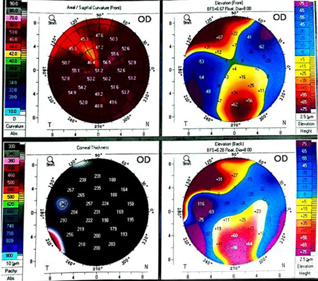
- OD: Kmax 56.7 D · SimK ≈ 53/54 D · astigmatism 9.3 D · thinnest pachymetry 154 μm
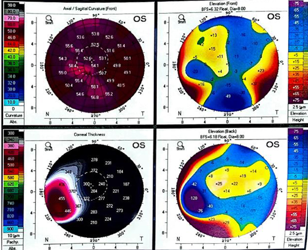
- OS: Kmax 60.2 D · SimK ≈ 53 D · astigmatism 3.1 D · thinnest pachymetry 140 μm
Pentacam 4-map (OD-OS): diffuse steepening, very low pachymetry, elevated posterior elevation.
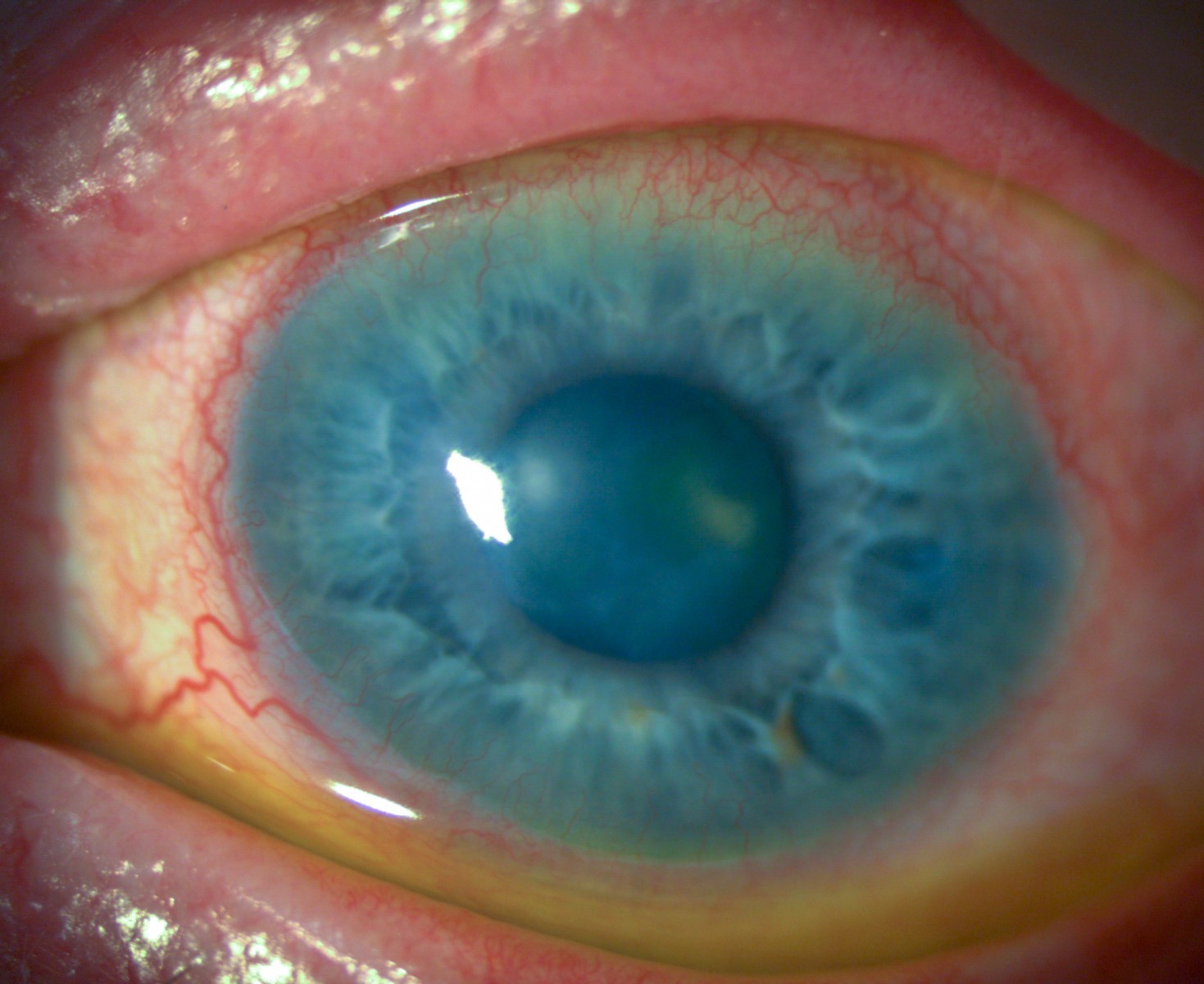
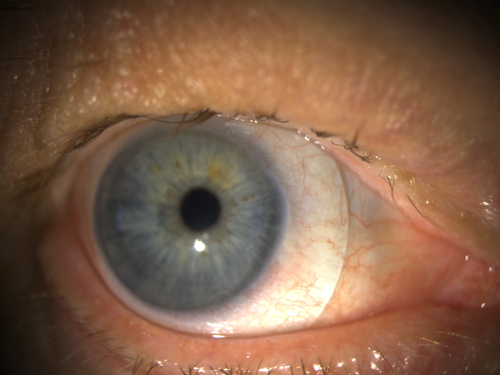
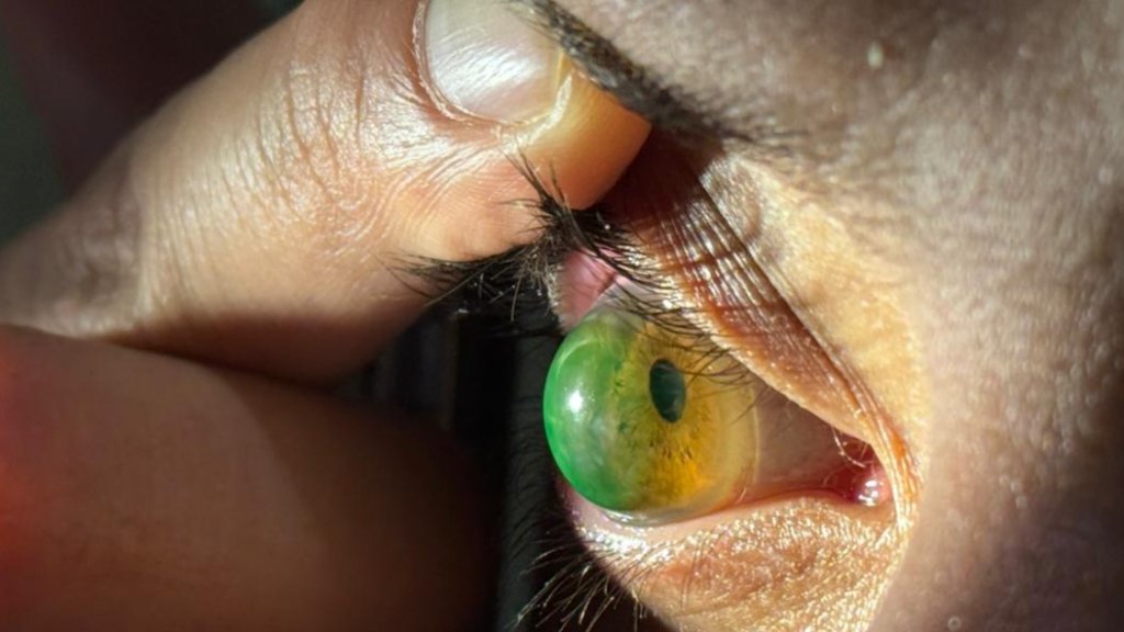
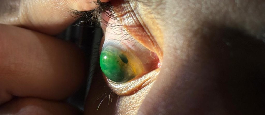
Globular corneal profile (OD) with the scleral lens in place, the whole-surface bulge is visible.
Corneo-scleral profilometry
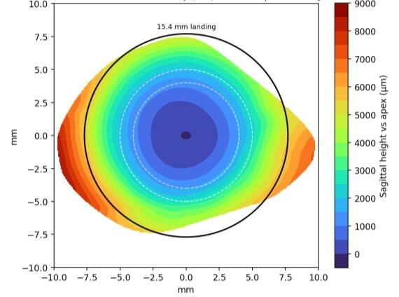
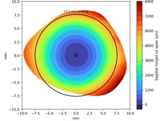
Both eyes were measured with corneo-scleral profilometry (ESP By Eaglet EYE), giving surface datasets of approximately 390,000 points (OD) and 436,000 points (OS). Central sagittal heights were high, consistent with the globular profile, and the corneo-scleral junction was asymmetric in both eyes.
Elevation map (OD) referenced to the apex, with the 15.4 mm landing ring
Elevation map (OS) referenced to the apex, with the 15.2 mm landing ring.
Lens design
Each lens was designed from the patient’s own corneo-scleral measurements rather than from standard fitting sets, using OraSight Freeform manufactured at VISIORA. The design objectives in each eye were to:
- Control sagittal depth (vault) over the cornea
- Land the lens on the sclera
- Move bearing away from the thin cornea
- Provide a regular optical surface over an irregular one
The two eyes were designed independently, since the ectatic profile differed between them.
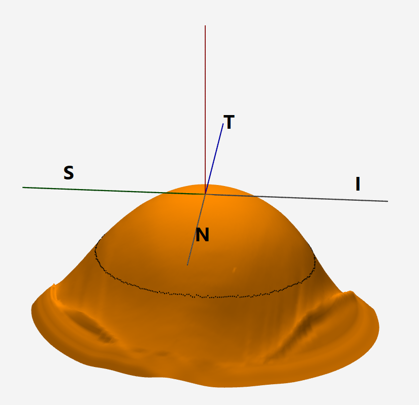
3-D reconstruction of the corneo-scleral surface, the globular dome and scleral transition.
| Parameter | OD | OS |
| Type | OraSight FREEFORM | OraSight FREEFORM |
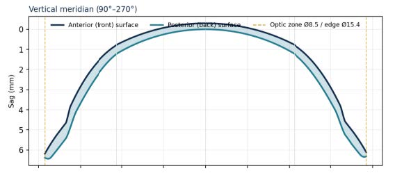
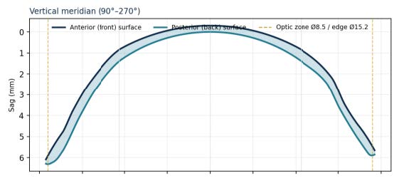
| Overall diameter | 15.4 mm | 15.2 mm |
| Base curve | 8.00 mm | 7.20 mm |
| Optic zone | 8.50 mm | 8.50 mm |
| Power | −5.00 D | −8.50 D |
| Central thickness | 300 μm | 300 μm |
| Average sag | 6,353 μm | 6,347 μm |
| Material – Dk | Optimum Extreme – 125 | Optimum Extreme – 125 |
Reconstructed lens cross-section (OD)
Reconstructed lens cross-section (OS)
A high-Dk material (Contamac Optimum Extreme, 125 Dk was used. A deep-vaulting scleral lens increases the oxygen-diffusion distance through the post-lens tear reservoir, and the high Dk helps compensate for this over a thin cornea.
Fitting and Settling
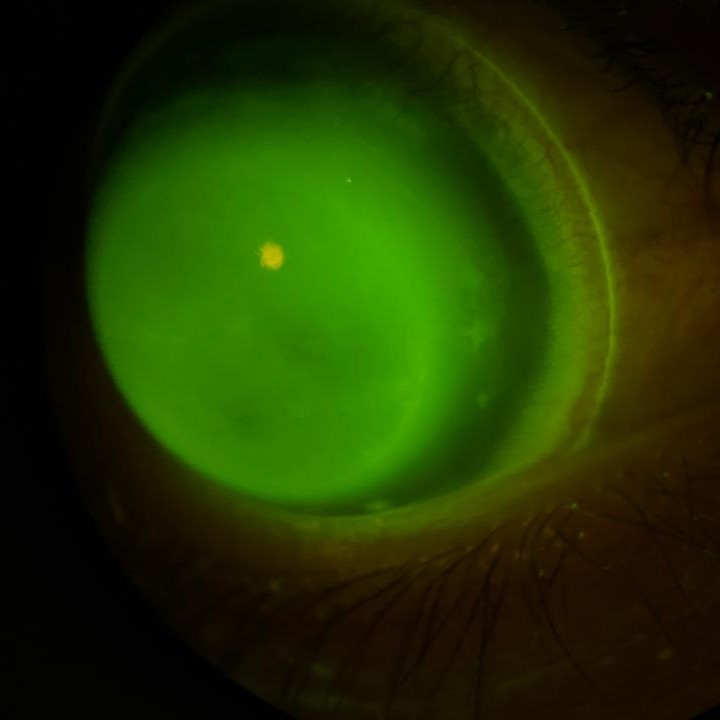
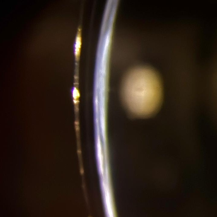
On-eye behavior was assessed over repeated sessions rather than from a single visit. The patient attended daily from Sunday to Wednesday, with supervised in-clinic wear of 2–3 hours per session. Scleral lenses settle over the first hours of wear, and the vault and landing were reviewed across sessions as they stabilized. On optical section, the lens-to-cornea clearance was approximately 250 μm over the apex, against a lens central thickness of 300 μm, with no central touch.
Fluorescein assessment showing the fluid reservoir vaulting the cornea, with clearance over the apex.
Optical (slit) section through the lens periphery: the scleral lens, the tear-fluid clearance, and the cornea are seen in sequence , showing lens thickness (300 μm) and a lens-to-cornea clearance of approximately 250 μm
Slit-lamp view (OD) with the scleral lens in place over the globular cornea; fluorescein highlights the tear reservoir and the whole-surface bulge beneath the lens.
Outcome
Best-corrected acuity with the OraSight Freeform lenses, compared with spectacles, was as follows. The fit was stable and comfortable with adequate vault control on examination.
| Eye | VA (Glasses) | VA (Freeform) |
| OD | 20/100 | 20/40 |
| OS | 20/80 | 20/40 |
The lens provided a stable optical surface and improved acuity in both eyes. It was not intended to treat the alternating esotropia, which was unchanged and is managed separately.
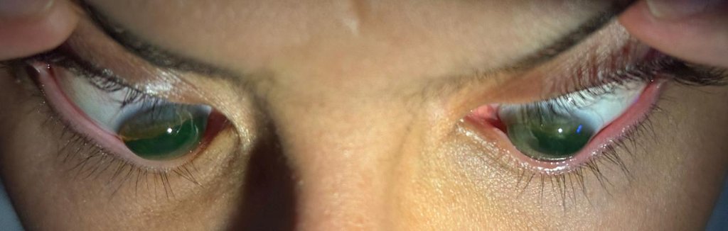
Both eyes with the OraSight Freeform scleral lenses in situ; the fluid-filled lenses vault each globular cornea.
Summary
In this bilateral keratoglobus case, freeform scleral lenses designed from each eye’s corneo-scleral measurements provided a stable, regular optical surface and improved best-corrected acuity to 20/40 in each eye (from 20/100 OD and 20/80 OS with spectacles). The design controlled the central vault, landed on the sclera, and kept bearing off the thin cornea. The lens addresses the optical surface only; it does not alter the underlying corneal thinning, and the patient remains under routine specialist follow-up.